This is used to acquire images of the areas that are in focus by applying the confocal principle. The resolution is high enough to present images of the structure in 3D, and of particular note, it is possible to observe cells in their living state.
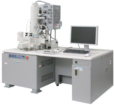
1. Resolution: 1.0nm guaranteed at 15kV or better
1.4nm guaranteed at 1kV or better
2. Magnification: x20 to x8000,000
3. Electron optics:
1) Electron gun: Cold or Schottky type field emission electron gun
2) Accelerating voltage: 0.1 to 30kV or wider
3) Lens system: 3-stage electromagnetic lens, Zoom reduction type
4) Objective lens aperture: 4 holes or more user selectable movable aperture
Aperture Heating function should be built-in.
5) Stigmator coil: Octopole electromagnetic system (X, Y)
6) Detector: Upper and Lower Secondary Electron Detector should be built-in.
It enables high-resolution image observation and composition analysis of micro/nano particles, bacteria and cells at low voltage, It is also used in the analysis of micro/nano particles such as gold, iron, and silica polymer and analysis of bacteria and animal-derived cells.
ⓒ KIMIRo. All rights reserved.