* This equipment allows examination of the morphology of the structure deposited with the drug of medical micro/nanorobots using low magnification but is also suitable for verifying the in vitro efficacy of medical micro/nanorobots, as it is also possible to check the morphology of each organelle inside a cell.
* It is capable of live cell imaging for observation of the changes in cancer cells caused by deposited drugs as well as robotic movement in real time, making it an essential piece of equipment for verifying the n vitro efficacy of micro/nanorobots.
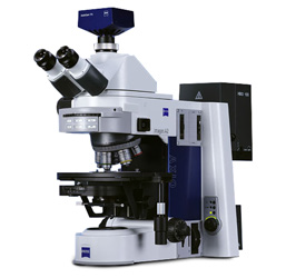
*Microscope: inverted stand, Manual stage with insert for slides, dish and multi-well plates
*Zeiss HBO arc lamp and power supply,
*Filter cubes: 1.DIC 2.DAPI, 3.CY5, 4.Green, 5.YFP, 6.Red, 7.CFP, 8.CY7
*Objectives: 5x/0.16, 10x/0.3, 20x/0.5, 40x/0.75, 63x/1.40, 100x/1.4
*Camera: Hamamatsu Orca ER digital camera & controller
Based on the principle that fluorescence occurs when UV light with short wavelengths is shined on the sample, the sample is treated with fluorescent material for observation. It is mainly used for genetic diagnosis or immunoassay of the sample under observation.
ⓒ KIMIRo. All rights reserved.